Introduction: Brain tumors, characterized by abnormal cellular proliferation within the brain or its surrounding structures, present significant medical challenges, including neurological impairments and potentially fatal consequences. Although certain brain tumors originate from genetic predispositions or unidentified etiological factors, accumulating evidence indicates that modifiable elements—such as lifestyle patterns, environmental exposures, and proactive healthcare measures—may contribute to reducing tumor risk. Given the often-limited curative interventions available for many brain tumor types, understanding preventive strategies is essential in minimizing disease incidence.
This paper aims to examine the scientific foundation of brain tumor prevention by evaluating modifiable risk determinants, including dietary habits, radiation exposure, chemical contaminants, and overall lifestyle behaviors. Through a comprehensive analysis of current research, this study seeks to offer evidence-based preventive recommendations for individuals striving to lower their susceptibility to brain tumors. Additionally, it underscores the critical role of early detection and preventive medical interventions in enhancing long-term neurological health outcomes.
What are the signs of a brain tumor?
The presence of a brain tumor does not always result in noticeable symptoms, particularly in cases where the tumor is small and does not exert significant pressure on surrounding brain structures. The clinical manifestations of a brain tumor are largely influenced by its size, type, and anatomical location within the brain.
How I knew I had a brain tumor? Common symptoms associated with brain tumors include:
- Headaches, which may be more intense in the morning or disrupt sleep.
- Seizures, occurring as a result of abnormal electrical activity in the brain.
- Cognitive and language impairments, leading to difficulties in thinking, speaking, or comprehending language.
- Personality and behavioral changes, which may manifest as mood disturbances or altered social interactions.
- Motor deficits, such as weakness or paralysis affecting one side of the body.
- Impaired coordination and balance, potentially causing dizziness or instability.
- Visual disturbances, including blurred or double vision.
- Auditory dysfunction, such as hearing loss or tinnitus.
- Sensory abnormalities, including facial numbness or tingling sensations.
- Gastrointestinal symptoms, such as nausea and vomiting, often due to increased intracranial pressure.
- Cognitive confusion and disorientation, which may impact memory and spatial awareness.
Given the potential severity of these symptoms, individuals experiencing persistent neurological disturbances should seek prompt medical evaluation to determine the underlying cause and initiate appropriate management strategies.
How common are brain tumors?
Brain tumors encompass a diverse group of over 100 distinct primary tumor types, each characterized by unique clinical manifestations, treatment approaches, and prognostic outcomes. Unlike many other malignancies, brain tumors frequently lead to profound and lasting physical, cognitive, and psychological impairments, significantly affecting patients’ quality of life. Despite extensive research efforts, survival rates for brain cancer have shown limited improvement over recent years, whereas notable advancements have been made in the prognosis of many other cancer types. Brain tumor age statistics indicate that the incidence and prevalence of brain tumors vary significantly across different age groups, with certain types being more common in pediatric populations and others predominantly affecting older adults.
According to the American Cancer Society, projections for brain and spinal cord tumors in the United States for 2025 include both adult and pediatric cases. It is estimated that approximately 24,820 malignant tumors of the brain and spinal cord will be diagnosed (14,040 in males and 10,780 in females), with actual incidence rates being substantially higher if benign tumors were included. Additionally, around 18,330 individuals (10,170 males and 8,160 females) are expected to succumb to these malignancies.
The lifetime risk of developing a malignant brain or spinal cord tumor is estimated to be less than 1%, with variations based on sex. While women exhibit a slightly higher overall risk of developing brain or spinal cord tumors, men are more likely to develop malignant forms of these tumors. This sex-related difference is attributed to the varying prevalence of specific tumor subtypes, such as meningiomas, which are more frequently observed in females. Understanding these epidemiological trends is critical for enhancing early detection strategies, optimizing treatment protocols, and ultimately improving patient outcomes.
Brain cancer survival rate
The 5-year survival rate for brain cancer is approximately 35–36%, though this figure varies significantly based on the tumor type and the patient’s age. Glioblastoma, the most prevalent malignant brain tumor, exhibits a notably lower 5-year survival rate, estimated at approximately 5–6%.
How fast do brain tumors grow?
The time required for a brain tumor to develop, from its origin to the point when symptoms begin to manifest, is influenced by various factors, particularly the tumor’s type and aggressiveness. The rate of growth and symptom onset can differ depending on the nature of the tumor. More aggressive tumors generally grow faster, while less aggressive ones may develop over a longer period. In the case of glioblastomas, which are among the most common and aggressive forms of brain cancer, their ability to remain undetected by the immune system often leads to delayed diagnosis and worsened prognosis.
The term glioma is used to describe primary tumors that originate in the brain tissue itself. Gliomas account for approximately 33% of all brain tumors. In contrast, many brain tumors, especially benign ones, grow slowly and may not necessitate immediate intervention. Some benign tumors can be safely monitored for months or years, with surgical removal only being considered when necessary. However, malignant tumors are typically more aggressive and require prompt treatment due to their rapid growth and potential for harm.
What causes brain tumors?
The precise biological mechanisms that drive the transformation of normal cells into tumor cells remain incompletely understood. However, it is widely accepted that brain tumor formation results from a complex interplay between genetic predisposition and environmental influences. Identified risk factors include metastasis from primary malignancies in other organs, inherited genetic disorders that promote excessive cell proliferation, and exposure to ionizing radiation.
Are brain tumors genetic?
Scientific evidence suggests that brain tumors arise due to genetic mutations affecting key regulatory genes within cellular chromosomes. These mutations disrupt essential cellular processes, including cell growth, division, and apoptosis (programmed cell death). Genetic alterations in brain cells can lead to aberrant signaling pathways that override normal regulatory mechanisms, resulting in uncontrolled cellular proliferation and prolonged survival. The accumulation of these genetically altered cells leads to tumor formation, which can exert pressure on surrounding brain structures and impair neurological functions.
Several inherited genetic mutations have been linked to an increased susceptibility to brain tumors. Hereditary syndromes, accounting for approximately 5% to 10% of brain tumor cases, include Neurofibromatosis type 1 (NF1 gene), Neurofibromatosis type 2 (NF2 gene), Turcot syndrome (APC gene), Gorlin syndrome (PTCH gene), Tuberous sclerosis complex (TSC1 and TSC2 genes), and Li-Fraumeni syndrome (TP53 gene). These conditions disrupt tumor-suppressor pathways, predisposing affected individuals to tumor development.
Environmental factors
In addition to genetic factors, environmental exposures significantly contribute to tumorigenesis. High-dose ionizing radiation from medical treatments or occupational exposure can induce DNA damage, increasing the likelihood of oncogenic mutations. In certain cases, environmental carcinogens alone may be sufficient to trigger genetic alterations that initiate tumor formation.
Understanding the genetic and environmental factors contributing to brain tumor development is crucial for advancing research in prevention, early diagnosis, and targeted therapeutic interventions. By identifying at-risk populations and mitigating environmental hazards, efforts can be directed toward reducing brain tumor incidence and improving patient outcomes.
Statistical analysis of tumor growth has revealed that the median specific growth rate of brain tumors is 1.4% per day, with an equivalent volume doubling time of about 49.6 days. Research into different tumor growth models has shown that the Gompertzian and linear radial growth models provide the best statistical fit, whereas the exponential growth model is less accurate for describing brain tumor growth dynamics.
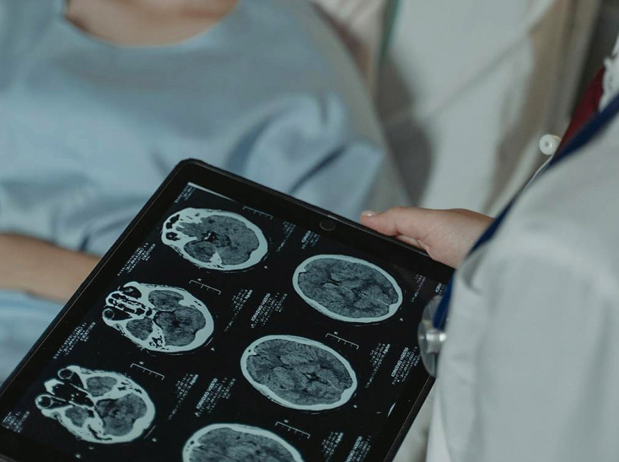
Brain tumour detection
The diagnosis of a brain tumor necessitates a series of specialized assessments and imaging techniques to evaluate neurological function and detect potential abnormalities. A comprehensive neurological examination is conducted to assess sensory and motor functions, including vision, hearing, balance, coordination, strength, and reflexes, aiding in the localization of neurological deficits. Imaging modalities play a crucial role in tumor detection, with computed tomography (CT) scans serving as an initial diagnostic tool to identify structural abnormalities, while magnetic resonance imaging (MRI) provides high-resolution images for more detailed evaluation. Advanced MRI techniques, such as functional MRI, magnetic resonance spectroscopy, and perfusion MRI, offer additional insights into tumor characteristics and vascular properties, facilitating precise diagnosis and treatment planning. Positron emission tomography (PET) scans, employing radioactive tracers, can detect metabolically active tumors, though their efficacy is limited for indolent or benign neoplasms.
When a brain tumor is suspected, histopathological confirmation through biopsy is often required. This procedure can be performed intraoperatively during surgical tumor resection or via stereotactic needle biopsy in cases where surgical intervention poses significant risks or when the tumor is in an anatomically challenging location. While biopsies are associated with potential complications, including hemorrhage and damage to adjacent brain structures, they provide critical diagnostic information regarding tumor histology, grading, and genetic mutations. Molecular and histological analyses of biopsy specimens enable the classification of the tumor as benign or malignant, as well as an assessment of its proliferative potential, thereby informing individualized therapeutic strategies.
Can a CT scan detect brain tumor?
A brain tumor diagnosis often begins with a neurological examination, followed by advanced imaging techniques such as magnetic resonance imaging (MRI) and computed tomography (CT) scans. MRI is particularly effective in producing high-quality images of soft tissues and blood vessels, making it an essential tool for detecting brain tumors. However, a CT scan can offer more detailed images of the bone structures surrounding the tumor, such as the skull and spine, providing valuable information for diagnosis. Additionally, CT scans are advantageous when MRI is not suitable, such as in patients with pacemakers, as MRI’s magnetic fields can interfere with pacemaker function. While the prospect of receiving a CT scan might cause anxiety, it is a reliable diagnostic method for brain tumors and plays a critical role in developing an appropriate treatment plan.
How accurate is CT scan for brain tumor?
While CT scans are highly effective in detecting large brain tumors, brain hemorrhages, and structural abnormalities, they may not be sufficient for identifying smaller or low-grade tumors, inflammation of the meninges, or certain soft tissue abnormalities.
Accuracy
Computed tomography (CT) scans serve as a reliable imaging modality for identifying large brain tumors, effectively ruling them out in diagnostic assessments. Additionally, CT imaging is particularly valuable for detecting intracranial hemorrhages, providing critical information in cases of acute brain injury. The detailed visualization of brain structures and tissues further enhances its diagnostic utility in neuroimaging.
Limitations
Despite their advantages, CT scans have certain limitations. They may fail to detect smaller or low-grade tumors and are not always effective in identifying meningeal inflammation. Furthermore, CT imaging does not offer the same level of soft tissue resolution as magnetic resonance imaging (MRI), making it less suitable for certain neurological conditions. Additionally, CT scans may not be capable of detecting all types of brain injuries or pathological conditions, necessitating the use of complementary imaging techniques when a more detailed evaluation is required.
How to prevent brain tumor?
While no definitive lifestyle choices have been scientifically proven to prevent brain tumors, adopting health-conscious ways to prevent brain tumor may contribute to overall well-being and lower the risk of malignancies. Several key brain tumor prevention strategies can support neurological and systemic health, potentially reducing cancer susceptibility:
- Nutritional Optimization – A diet rich in fruits, vegetables, and whole grains provides essential nutrients, supports immune function, and reduces systemic inflammation, which may help lower cancer risk.
- Regular Physical Activity – Engaging in consistent exercise promotes healthy body weight, enhances cardiovascular health, and improves cerebral blood flow, which may contribute to reducing the likelihood of tumor development.
- Avoidance of Carcinogenic Substances – Eliminating tobacco use and limiting excessive alcohol consumption are crucial in reducing exposure to harmful compounds linked to cancer formation, including those affecting the central nervous system.
- Minimization of Environmental Exposures – Reducing contact with hazardous chemicals, radiation, and other environmental risk factors may decrease potential DNA damage, which can contribute to tumor development.
Although direct preventive measures for brain tumors remain uncertain, adopting these health-promoting strategies may contribute to overall cancer risk reduction and improved neurological health.
What foods prevent brain tumor?
While no single food can prevent diseases like cancer, certain foods, often called “superfoods,” have shown potential in reducing cancer risk due to their cancer-fighting properties. A brain tumor prevention diet focuses on nutrient-rich foods high in antioxidants, phytochemicals, and anti-inflammatory compounds, which may help reduce oxidative stress and support overall brain health. Nutritionist Pooja Malhotra highlights the health benefits of foods rich in phytochemicals and antioxidants, which are crucial in cancer prevention.
Foods to prevent brain tumors:
- Beans: A nutrient-dense alternative to meat, beans may reduce cancer risk and recurrence, according to studies. Eating them a few times a week is beneficial.
- Berries: Rich in antioxidants, berries like strawberries and blueberries protect against cancer by neutralizing harmful radicals that may cause cell mutations.
- Tomatoes: The antioxidant lycopene, responsible for tomatoes’ red color, helps protect against cancer, as supported by research.
- Cruciferous Vegetables: Vegetables such as broccoli, spinach, and kale are known for their cancer-protective properties and overall health benefits.
- Turmeric: A staple in South Asian cuisine, turmeric contains anti-inflammatory properties and enhances immunity, potentially reducing cancer risks.
- Flax Seeds: These nutrient-packed seeds may slow cancer cell growth and could even help eliminate them.
- Nuts: Regular consumption of nuts, particularly almonds, is linked to reduced risks of brain tumors and other cancers.
- Garlic: The compound allicin in garlic has demonstrated anti-cancer effects, including the ability to kill cancer cells.
- Citrus Fruits: Rich in vitamin C, citrus fruits like oranges and lemons protect against cancer by helping prevent cellular mutations caused by UV rays.
- Fatty Fish: High in omega-3 fatty acids, fatty fish may reduce cancer risk, contrasting with the harmful effects of red meats.
Incorporating these superfoods into your diet, along with regular exercise, can significantly impact overall health and potentially lower cancer risks.
Can exercise prevent brain tumor
Although physical activity does not directly prevent brain tumors, brain tumor prevention exercise offers significant benefits for individuals diagnosed with or recovering from brain cancer by enhancing physical function, mental well-being, and overall quality of life. Exercise has demonstrated positive effects across various cancer types and is increasingly recognized as a supportive intervention for glioblastoma (GBM) and other brain tumors.
As a safe and viable adjunctive therapy, structured exercise programs can contribute to improved quality of life in patients with brain tumors, including those with GBM. Additionally, physical activity may influence tumor progression through several biological mechanisms, such as:
- Enhancing vascularization, Tissue perfusion
- modulating immune function,
- Altering tumor metabolism
- Mediating muscle–tumor interactions
These findings suggest that integrating exercise into brain tumor management could provide therapeutic advantages beyond conventional treatments.
How to prevent brain tumor at home
Although there is no definitive way to prevent brain tumors, adopting a healthy lifestyle can help reduce risk factors and promote brain health. A nutrient-rich diet with antioxidants, omega-3 fatty acids, and neuroprotective foods like turmeric and green tea can combat inflammation and oxidative stress. Limiting radiation exposure from mobile devices and environmental toxins, engaging in regular exercise, maintaining adequate sleep, and managing stress through meditation or yoga further support brain function. Avoiding carcinogens like tobacco and alcohol, strengthening the immune system with vitamin D and hydration, and staying vigilant with regular health check-ups—especially for those with a family history—are additional proactive measures to promote overall neurological well-being.
How to prevent brain tumor recurrence
Long-term studies have demonstrated that nearly half of surgically excised brain tumors reemerge within two decades, emphasizing the necessity for ongoing surveillance. Despite the potential for recurrence, patients can maintain a normal quality of life through consistent monitoring. Several key determinants influence the likelihood of meningioma recurrence following surgical intervention. These include:
- Completeness of tumor resection
- Tumor grade
- size
- Location
- Presence of multiple tumors
Brain tumor prevention recurrence involves a combination of medical treatments, lifestyle modifications, and regular monitoring. While complete prevention is not always possible, the following strategies can help reduce the risk:
- Adjuvant Therapies
- Regular Monitoring
- Stress and Immune System Management
- Genetic and Molecular Research
Brain tumor vaccine
The treatment of brain tumors presents numerous challenges, including the risk of recurrence despite advanced medical interventions. The immune system, composed of specialized cells, antibodies, and organs, plays a crucial role in defending the body against harmful pathogens. However, its ability to differentiate between normal and cancerous cells often requires external support.
Vaccine therapy is an emerging immunotherapeutic strategy designed to enhance the immune system’s ability to recognize and target brain tumor cells. This personalized approach involves extracting tumor tissue through a biopsy during surgery, which is then utilized to develop a patient-specific vaccine. Through a series of regular injections, the vaccine stimulates and strengthens the immune response, equipping the body to combat cancerous cells more effectively.
As research continues to advance, brain tumor vaccines represent a hopeful frontier in neuro-oncology, offering a personalized and potentially life-extending approach for patients facing the complexities of malignant brain tumors.
Brain tumor stories from survivors
Here are accounts of individuals who have successfully overcome brain tumors.
Mary Ann’s Story
Mary Ann Gunderman, a 73-year-old grandmother, experienced severe facial pain after being struck by an ocean wave during a vacation. She initially attributed the pain to a dental issue, as she had a similar but brief episode a year earlier. Despite undergoing dental procedures, her condition worsened, leading her to seek medical help. After being dismissed by an emergency room, she was finally diagnosed with trigeminal neuralgia by an oral surgeon. An MRI revealed a large tumor at the base of her skull, pressing against her trigeminal nerve and brainstem. She was referred to Dr. Jennifer Moliterno Gunel at Yale’s Smilow Cancer Hospital, where a team of specialists confirmed the diagnosis and recommended surgery to relieve the pressure while minimizing complications. Dr. Moliterno successfully removed almost the entire benign meningioma during an eight-hour operation, preserving Mary Ann’s hearing and facial function. Remarkably, she was walking within two days and discharged after four days with no need for further treatment. Over time, her facial numbness significantly improved, and she remained pain-free. Grateful for the expertise and compassion of the Yale medical team, she reflected on how the experience changed her perspective, emphasizing the importance of family, friends, and appreciating life’s moments. The ordeal reinforced her belief in the value of expert medical care and the support of loved ones, leaving her with a newfound appreciation for life. (19)
Caroline’s Story
During her freshman year at Michigan State University, the author’s life took an unexpected turn when a persistent sinus infection led to the incidental discovery of a brain tumor. An MRI revealed an unidentified lesion in her brain, prompting a consultation with Dr. Ian Lee at Henry Ford Health. Initially, they decided to monitor the lesion for two months before performing a biopsy, which confirmed the presence of a grade 2 astrocytoma glioma. Despite the daunting diagnosis, she remained committed to her education, scheduling surgery for January 8 while coordinating accommodations to continue her studies. The seven-hour operation was successful, and despite exhaustion, she quickly resumed her coursework, defying expectations about recovery time and pain tolerance. However, post-surgery pathology results revealed signs of a more aggressive tumor, necessitating 33 rounds of radiation and daily oral chemotherapy for seven weeks. Determined not to let cancer disrupt her life, she commuted daily between school and treatment, balancing rigorous academic demands with the physical toll of therapy. In a remarkable feat, she completed finals, moved out of her dorm, and celebrated the end of her treatments—all in the same week. Grateful for the unwavering support of her medical team, family, and friends, she reflected on her eight-month journey, during which she underwent two surgeries, endured months of treatment, and drove thousands of miles—all culminating in the triumphant defeat of her brain tumor. (19)
Debbie Gilbert-Gardner’s journey with glioblastoma multiforme began with a persistent headache that lasted three weeks, ultimately leading her to seek medical help at Henry Ford Health. Diagnosed with one of the most aggressive forms of brain cancer, she was quickly transferred to a specialized team led by Dr. Ian Lee. Due to the tumor’s deep location in her thalamus, traditional surgery was not an option, and she underwent a cutting-edge laser ablation procedure that successfully removed 85% of the tumor. Following surgery, Debbie received chemotherapy, radiation therapy, and utilized a wearable device called Optune, which employs tumor-treating fields to slow cancer progression. Through the unwavering support of her medical team and family, she faced the battle with resilience, determined to live life to the fullest. Throughout her treatment, Debbie found strength in her faith, family, and daily joys, such as babysitting her grandson and preparing for a long-awaited trip to the Bahamas. The experience transformed her perspective, making her appreciate life’s precious moments while letting go of negativity. She remains optimistic, embracing every day with gratitude and a renewed sense of purpose. Debbie continues to rely on Henry Ford’s advanced care, acknowledging the expertise of her doctors and the emotional support they provided. As she looks to the future, her goal is to see her grandchildren grow and enjoy the simple pleasures of life, all while advocating for perseverance and positivity in the face of adversity. (19)
Ken Gipfert, who historically avoided doctors due to a lack of health insurance, was diagnosed with Stage 4 cancer that had metastasized from his lung to his brain after experiencing a seizure at work. He underwent multiple surgeries, radiation treatments, and experimental procedures at Henry Ford Hospital and The Hermelin Brain Tumor Center. Despite the tumor recurring multiple times, advanced treatments such as laser ablation, awake craniotomy, and GammaTile implantation were used to control the disease. Ken’s resilience and faith remained strong despite losing some function in his left arm and facing complications like a small stroke. Five years after his initial diagnosis, Ken has defied the odds with remarkable stability following his latest surgery. His case highlights the importance of multidisciplinary cancer care, integrating neurosurgery, oncology, and advanced radiation techniques. Though his active lifestyle has changed, Ken remains grateful for what he can do and continues to maintain a positive outlook. Supported by Henry Ford’s ExCITE program, he focuses on regaining mobility and inspiring others. Despite the challenges, he remains determined, setting high expectations for his recovery, proving that faith and perseverance can make all the difference. (19)
Nick’s Story
Nick Petrykowski, a determined physical therapy aide and Michigan State University student, experienced recurring visual and auditory disturbances for months, initially mistaking them for panic attacks. After worsening episodes, he was diagnosed with a brain tumor in the right posterior parietal lobe. Neurosurgeon Dr. Jack Rock successfully removed the grade 2 pilomyxoid astrocytoma, and doctors opted against chemotherapy and radiation due to the tumor’s genetic characteristics and Nick’s strong health. His recovery was gradual, but he remained resilient, crediting his family, faith, and medical team for their support. Now accepted into medical school, Nick plans to specialize in neurology, using his experience to empathize with future patients. He advises others to seek timely medical attention and appreciate life’s small moments. (19)
Conclusion
The clinical presentation of brain tumors varies significantly depending on factors such as tumor location, size, growth rate, and histological type. Common symptoms include persistent headaches, seizures, cognitive and personality changes, motor and sensory deficits, and signs of increased intracranial pressure such as nausea and vomiting. The variability in symptoms often leads to delayed diagnosis, underscoring the importance of early recognition and neuroimaging for timely intervention. Advanced diagnostic modalities, including MRI, CT scans, and biomarker analysis, have improved the accuracy of tumor detection and classification. Given the complexity of brain tumor manifestations, a multidisciplinary approach involving neurology, oncology, and neurosurgery is essential for optimizing patient outcomes. Further research into molecular and genetic markers may enhance early detection and individualized treatment strategies, ultimately improving prognosis and quality of life for affected individuals.
Frequently Asked Questions
Here are Frequently Asked Questions about brain tumor:
What does a brain tumor feel like?
Headaches, seizures (fits) persistently feeling sick (nausea), being sick (vomiting) and drowsiness. mental or behavioral changes, such as memory problems or changes in personality.
What is the symptom for brain tumor?
Common symptoms include: headaches. Seizures (fits) persistently feeling sick (nausea), being sick (vomiting) and drowsiness. Mental or behavioral changes, such as memory problems or changes in personality. Progressive weakness or paralysis on one side of the body. Vision or speech problems.
How I found out my child had a brain tumor?
Some of the more common signs and symptoms of pediatric brain tumors include: Headaches, which may become more frequent and more severe. In children who don’t talk, a parent might notice that the child is more irritable than usual. Nausea and vomiting.
How to avoid brain tumor?
Unfortunately, you can’t prevent a brain tumor. You can reduce your risk of developing a brain tumor by avoiding environmental hazards such as smoking and excessive radiation exposure.
Does depo Provera cause brain tumors?
Emerging evidence now connects Depo-Provera to a serious health risk—intracranial meningiomas—slow-growing brain tumors that can lead to severe, life-altering health complications.
Can brain tumors be prevented?
Unfortunately, you can’t prevent a brain tumor. You can reduce your risk of developing a brain tumor by avoiding environmental hazards such as smoking and excessive radiation exposure.
Reference:
- https://pubmed.ncbi.nlm.nih.gov/29506669/
- https://pubmed.ncbi.nlm.nih.gov/32478924/
- https://pubmed.ncbi.nlm.nih.gov/34628433/
- https://pubmed.ncbi.nlm.nih.gov/30655533/
- https://pubmed.ncbi.nlm.nih.gov/25791797/
- https://pubmed.ncbi.nlm.nih.gov/32152825/
- https://pubmed.ncbi.nlm.nih.gov/39406603/
- https://pubmed.ncbi.nlm.nih.gov/32890023/
- https://pubmed.ncbi.nlm.nih.gov/38893253/
- https://www.thelancet.com/journals/laneur/article/PIIS147444220870111X/abstract
- https://www.nature.com/articles/s41571-019-0177-5
- https://www.thelancet.com/journals/lancet/article/PIIS0140-6736(03)12328-8/abstract
- https://onlinelibrary.wiley.com/doi/abs/10.1111/j.1742-1241.2001.tb11093.x
- https://journals.plos.org/plosone/article?id=10.1371/journal.pone.0314721
- https://www.nature.com/articles/s41568-022-00475-0
- https://link.springer.com/article/10.1186/s12943-022-01668-9
- https://www.nature.com/articles/s41571-021-00529-6
- https://academic.oup.com/comjnl/article-abstract/67/3/1126/7147953
- https://www.henryford.com/services/brain-tumors/patient-stories